Comprehensive Eye and Vision Examination
Periodic eye and vision examinations are an important part of preventive health care. Many eye and vision problems have no obvious signs or symptoms. As a result, individuals are often unaware that problems exist. Early diagnosis and treatment of eye and vision problems are important for maintaining good vision and eye health, and when possible, preventing vision loss.
A comprehensive adult eye and vision examination may include, but is not limited to, the following tests. Individual patient signs and symptoms, along with the professional judgment of the doctor, may significantly influence the testing done.
Patient History
A patient history helps to determine any symptoms the individual is experiencing, when they began, the presence of any general heath problems, medications taken and occupational or environmental conditions that may be affecting vision. The doctor will ask about any eye or vision problems you may be having and about your overall health. The doctor will also ask about any previous eye or health conditions of you and your family members.
Visual Acuity
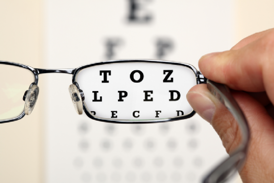
Reading charts are often used to measure visual acuity. Visual acuity measurements evaluate how clearly each eye is seeing. As part of the testing, you are asked to read letters on distance and near reading charts. The results of visual acuity testing are written as a fraction such as 20/40.
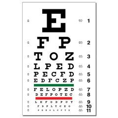
When testing distance vision, the top number in the fraction is the standard distance at which testing is done, twenty feet. The bottom number is the smallest letter size you were able to read. A person with 20/40 visual acuity would have to get within 20 feet of a letter that should be seen at 40 feet in order to see it clearly. Normal distance visual acuity is 20/20.
Preliminary Tests
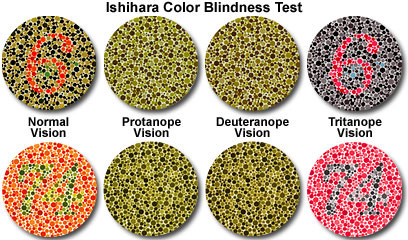
Preliminary testing may include evaluation of specific aspects of visual function and eye health such as depth perception, color vision, eye muscle movements, peripheral or side vision, and the way your pupils respond to light. In the Ishihara color plates below, can you see the number 6 and 74?
Protanope – Theses individuals cannot see the 6 and 74 plates in the first column due to a red-green color blindness deficiency. Basically, they cannot distinguish well the red 6 and green 74 below and, therefore, only see a green plate.
Deuteranope – Are either green color weak or green color blind. Again, they would have difficulty viewing the 6 and 74.
Tritanope – Although called “Blue-Yellow” color blindness, these individuals have difficulty with blue and green colors.
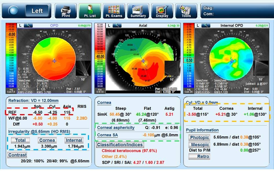
Keratometry
This test measures the curvature of the cornea, the clear outer surface of the eye, by focusing a circle of light on the cornea and measuring its reflection. This measurement is particularly critical in determining the proper fit for contact lenses and evaluating the cornea for corneal diseases like Keratoconus shown below.
Refraction
Determining refractive error with a phoropter and retinoscope is conducted to determine the appropriate lens power needed to compensate for any refractive error (nearsightedness, farsightedness, or astigmatism). Using an instrument called a phoropter, your optometrist places a series of lenses in front of your eyes and measures how they focus light using a hand held lighted instrument called a retinoscope. The doctor may choose to use an automated instrument that automatically evaluates the focusing power of the eye. The power is then refined by patient’s responses to determine the lenses that allow the clearest vision.
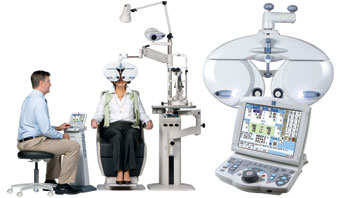
This testing may be done without the use of eye drops to determine how the eyes respond under normal seeing conditions. In some cases, such as for patients who can’t respond verbally or when some of the eyes focusing power may be hidden, eye drops are used. The drops temporarily keep the eyes from changing focus while testing is done.
Eye Focusing, Eye Teaming, and Eye Movement Testing
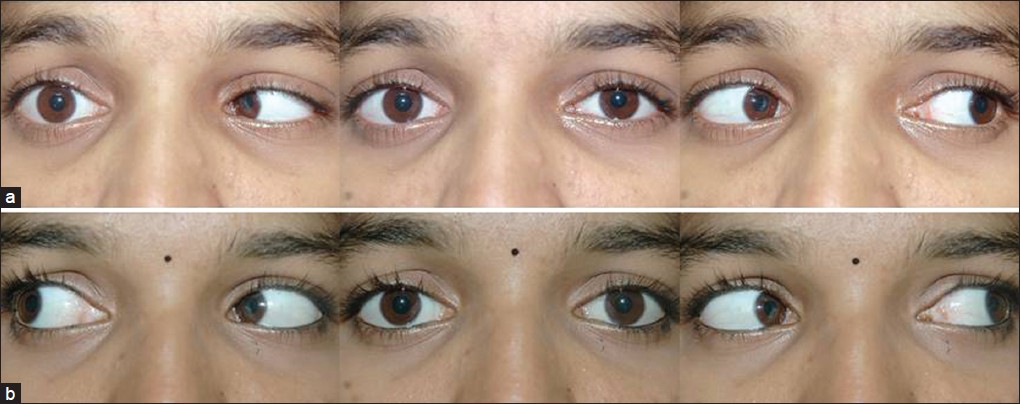
Assessment of accommodation, ocular motility and binocular vision determines how well the eyes focus, move and work together. In order to obtain a clear, single image of what is being viewed, the eyes must effectively change focus, move and work in unison. This testing will look for problems that keep your eyes from focusing effectively or make using both eyes together difficult. Below you will see an abducens (cranial nerve 6) nerve palsy. This patient, example on top, is unable to move his right eye out towards his ear(look to his right) due to a palsy on cranial nerve 6.
Eye Health Evaluation
Tonometry measures eye pressure. Elevated pressure in the eye signals an increased risk for glaucoma. External examination of the eye includes evaluation of the cornea, eyelids, conjunctiva and surrounding eye tissue using bright light and magnification.
Measurement of pressure within the eye (tonometry) is performed. Normal eye pressures range from 10 to 21 millimeters of mercury (mm Hg), averaging about 14 to 16 mm Hg. Anyone with eye pressure greater than 22 mm Hg is at an increased risk of developing glaucoma, although many people with normal pressure also develop glaucoma. Glaucoma is tricky as it has no signs and symptoms. Many patient who are diagnosed with glaucoma had no idea that they had it. Even patients with perfect vision are affected equally as those who wear glasses.
Evaluation of the lens, retina and posterior section of the eye may be done through a dilated pupil to provide a better view of the internal structures of the eye. Here are a few examples of what optometrists can see during a routine dilated eye examination.
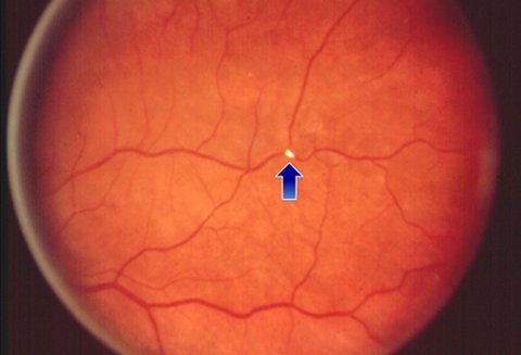
Cholesterol(Hollenhorst plaque)plaque in the arterial blood vessel of the back of the eye
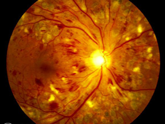
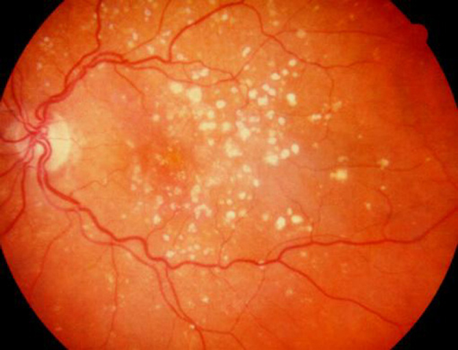
Uncontrolled Diabetes can lead to vision changes and bleeding in the back of the eye, Diabetic Retinopathy
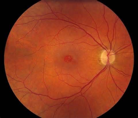
Elevated blood pressure can lead to blood vessel changes in the eye, Hypertensive Retinopathy
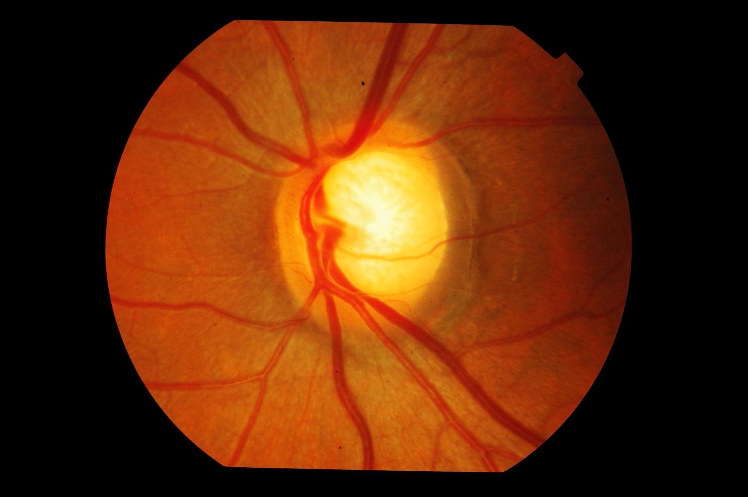
Glaucoma – Optic nerve head changes resulting in cupping
Age Related Macular Degeneration can lead to central vision loss/distortion
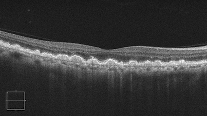
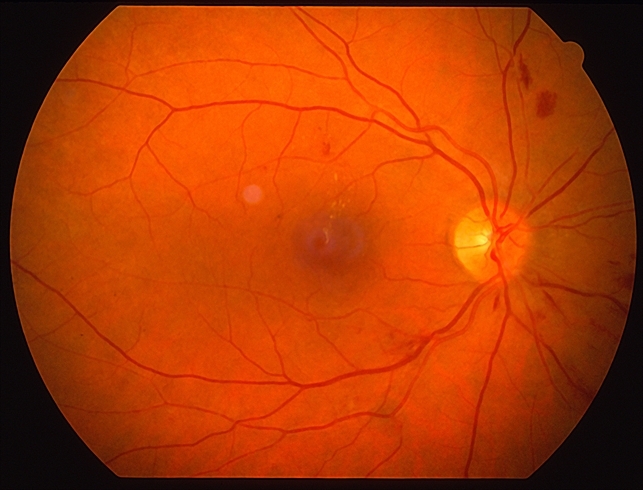
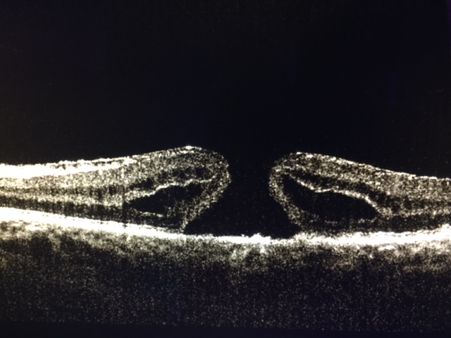
Full thickness (Stage III) Macular Hole. As we age, the vitreous slowly shrinks and pulls away from the retinal surface. However, if the vitreous is firmly attached to the retina when it pulls away, it can tear the retina and create a macular hole.
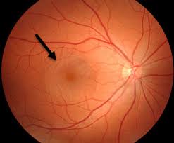
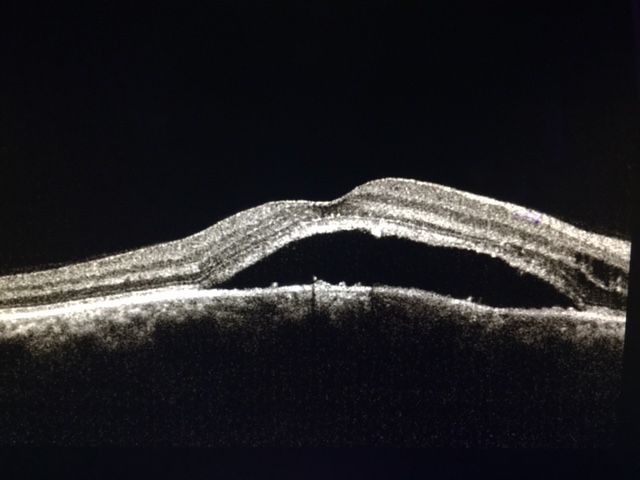
Excess Cortisol/Corticosteroid intake or stress can lead to leakage of fluid under the retina known as Central Serous Retinopathy
Supplemental testing
Additional testing may be needed based on the results of the previous tests to confirm or rule out possible problems, to clarify uncertain findings, or to provide a more in-depth assessment.
At the completion of the examination, your optometrist will assess and evaluate the results of the testing to determine a diagnosis and develop a treatment plan. He or she will discuss with you the nature of any visual or eye health problems found and explain available treatment options. In some cases, referral for consultation with, or treatment by, another optometrist or other health care provider may be indicated.
If you have questions regarding any eye or vision conditions diagnosed, or treatment recommended, don’t hesitate to ask for additional information or explanation from your doctor.